
Renaissance/Enlightenment (1600’s-1800’s)
38 The Beginning of Anatomical Study
Alex Fischer; Alexandra Harrington; and Ashlyn Miles
introduction
The early development of anatomical study dates back to around 300 BC with the work of Herophilus and Erasistratus in Alexandria. These individuals made a significant shift in medicine from speculation to empirical and observational reasoning. Galen, a physician in the Roman Empire, made advancements from the work of Herophilus and Erasistratus around the 2nd century AD. Galen furthered anatomical understanding through his work based on animal dissections. These three individuals played key roles in the early understanding and advancements in anatomical study.
The Renaissance was a time of rebirth and intellectual growth that ignited curiosity and discovery across Europe. While some were creating new mechanical monsters, like the printing press or the microscope, others turned their attention to the previously unexplored human anatomy. The works of Leonardo da Vinci, Andreas Vesalius, and many other brilliant individuals during the Renaissance sparked a shift toward anatomy and dissection in society and advanced medicines, influencing countless works of art and diagrams that are still praised today. As we will see, these discoveries deepened society’s understanding of the scientific world around us, and in turn, set new standards of medical understanding, education, and care for people around the globe.
COnnection to Science, Technology, and society
Herophilus, Erasistratus, Galen, and Susini connect to Science, Technology, and Society as their contributions laid the groundwork for the beginning of anatomical study. Herophilus and Erasistratus’ observational work through dissections marked a shift from a theory-based understanding to observational reasoning, influencing the scientific method that would later become crucial to modern science. Galen built upon the findings of Herophilus and Erasistratus to influence medical knowledge for centuries. Galen not only impacted scientific understanding, but he also influenced technological advancements in medicine. The work of Herophilus, Erasistratus, and Galen made profound societal impacts with contributions to the development of medicine, improved public health, and sparked philosophical debates about anatomy. These early anatomists helped bridge the gap between ancient theoretical understandings and scientific innovations that would emerge in later centuries, emphasizing the relationship between science, technology, and society.
The Renaissance marked a deepening of scientific understanding of the human body like never before. Science, Technology, and Society (STS) is defined by Harvard University as the explores “what difference it makes to human societies that we, collectively, are producers and users of science and technology” (What Is STS?, 2024). This newfound interest in anatomy spurred the creation of new technologies, such as dissection tools, diagrams, and medicines. Societal impacts were profound, ranging from public dissections that prompted the spread of scientific knowledge to the undeniable influence on art. The study of anatomy during the Renaissance was paramount to the advances in science, technology, and society of the time.
Herophilus and Erasistratus
Herophilus of Alexandria is known as the Father of Anatomy and studied the first ever human cadaver (Bay, 2010). He is best known for being the first to systematically dissect the human body, an achievement that was revolutionary at the time. His contributions to anatomical study predates even Andreas Vesalius, who is often considered the founder of modern anatomy. Despite the taboo surrounding the desecration of human bodies, Herophilus and his contemporary, Erasistratus, conducted vivisections, a practice that led to controversy but also provided profound insights into human physiology. Herophilus made significant observations in a variety of bodily systems, including the brain, eye, liver, reproductive organs, and nervous system. He was the first to differentiate between nerves and blood vessels and to identify the brain as the “seat of intellect.” He also made notable advancements in understanding the cardiovascular and digestive systems, as well as the structure of the reproductive organs. Herophilus’ work laid the foundation for centuries of anatomical study and was instrumental in shaping the future of medicine, despite the eventual cessation of human dissection for nearly two millennia.
Leonardo Davinci
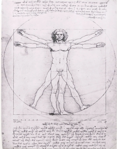
Leonardo da Vinci is possibly the most well-known figure of the Italian Renaissance. He is typically remembered for his artwork, but he also made great strides in the world of anatomical discovery. During his apprenticeship in Verrocchio’s workshop, da Vinci became entranced by the workings of the human body (Heydenreich, 2018). This fascination traveled with him to the city of Milan, where he dove deeper into the skeletal and muscular systems that composed the body. To do this, da Vinci conducted numerous dissections, eventually making his way into hospitals in Florence and Rome, where he ultimately dissected up to 30 corpses in his lifetime (Heydenreich, 2018). As we will discuss later, dissections were a very new idea at this time, and often considered taboo. Da Vinci’s willingness to break societal norms for the sake of scientific discovery is what allowed him to be the scientist and artist he was. Much of his art incorporated the human figure. Pictures like the famous Vitruvian Man and his sketches of the human fetus and a man seen below are perfect examples of how his artwork was molded by his scientific mind (da Vinci, c. 1490, as cited in Heydenreich, 2018). These works of art are lasting and still have an impact on modern society today. Da Vinci’s innate curiosity and revolutionary approach to anatomy laid the foundations that continue to influence modern medical understanding, but he was not the only one.
Andreas Vesalius
Andreas Vesalius does not receive as much recognition as deserved but is an equally influential figure in the development of anatomical sciences during the Renaissance. Vesalius was raised in the scientific world, with his family consisting of physicians and pharmacists. He attended the University of Paris, where he began his own path of dissection, initially on animals. As Florkin mentions in Britannica, Vesalius also “devoted much of his time to a study of human bones, at that time easily available in the Paris cemeteries” (Florkin, 2018). Vesalius’s extreme desire to learn drove him to the lengths of unearthing human corpses to study what had never been studied before. He later earned his doctoral degree in medicine from the University of Padua and began teaching his anatomical findings through public demonstrations. Through these dissections, Vesalius developed his own theories about the human body that contradicted the predominant theories of his time. He realized that the theories of Galen, which were based solely on animal dissections, were incorrect and set out to correct them (Florkin, 2018). Vesalius wrote and published the first comprehensive book on Western anatomy, De humani corporis fabrica, revolutionizing the field and cementing his claim as the “Father of Modern Anatomy” (Florkin, 2018). His work laid the foundation for modern anatomy and transformed the scientific understanding of the human body, paving the way for more advanced medical practice.
Galen
Claudius Galenus, or Galen, is known as the Father of Systematic Medicine with his significant contributions to anatomical and physiological concepts that shaped societal understanding for the next 15 centuries (Pasipoularides, 2014). Galen’s extensive writings of over 600 treatises covered many medical topics, although only a fraction of them survive today. His work became the ultimate authority in medicine for over a millennium. His anatomical theories remained largely unchallenged until the 17th century. Galen was a master of experimental methods, emphasizing the importance of directly testing nature and using empirical evidence to validate medical knowledge. His groundbreaking research spanned many disciplines, including embryology, neurology, myology, and urology, and he was the first to use the pulse as an indicator of illness. Galen’s anatomical concepts were pivotal in advancing the understanding of the body’s systems, though some of his theories, such as his belief in the communication between veins and arteries through invisible “anastomoses,” were later proven incorrect. Despite his errors, his ideas on the organization of the venous, arterial, and nervous systems and their respective functions in distributing natural, vital, and animal spirits profoundly influenced the development of medicine, including William Harvey’s groundbreaking work on the circulatory system. Galen’s contributions to both clinical practice and pharmacology, combined with his ethical approach to patient care, solidified his legacy as one of the most influential figures in the history of medicine.
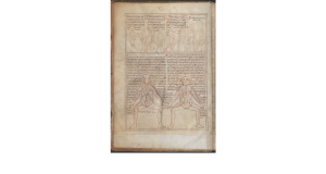
Andreas Galen was a Greek physician, philosopher, and anatomist living in Rome in the second century A.D.. He had a significant influence on the practice and study of medicine that has been handed down over many generations, still feeling his impacts today. One of his most profound contributions was the introduction to the five major anatomical systems of the human body, including the musculoskeletal, nervous, cardiovascular, respiratory, and digestive systems. Although the origins are not fully known, the “Five-Figure Series”, as named by medical historian Karl Sudhoff, diagrams the bones, nerves, arteries, muscles, and veins as described by Galen’s teachings (Laurenza, 2012). The school of Alexandria incorporated these diagrams into their teachings in the ‘sixteen books of Galen’ (French, 1984).
Clemente Susini
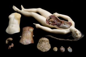
Clement Susini, an Italian sculptor, is best known for his Anatomical Venus, also referred to as “The Demountable Venus” (Susini, c. 1790, as cited in Ebenstein, 2012). He created this hyperreal, life-sized, dissectable sculpture of a woman to establish a new way to teach human anatomy. This sculpture could be disassembled into several parts and explored to better understand the female anatomy. The final removal revealed a fetus in a curled position in the wax woman’s womb, allowing for instruction on not only the interior organs but also those required for sexual reproduction. (Ebenstein, 2012). Susini also created sisters for this original Anatomical Venus, known as The Slashed Beauty and The Dissected Graces. Cadavers were not readily available due to medical ethics as well as their tendency to decompose quickly, so these wax sculptures became essential for instruction without dissection, as well as an attraction and wonder to a wide audience.
Furthermore, these structures have established a connection, rather than a divide, on some fine lines that define life as we know it. As shared by artist Joanna Ebenstein, Susini’s wax women “confound, troubling the edges of our neat categorical divides: life and death, science and art, body and soul, effigy and pedagogy, spectacle and education, kitsch and art” (2012). Susini added a divine element, simultaneously teaching female anatomy while also dramatizing and romanticizing it.
Impact on sugEry and medicine

Without the anatomical work of Vesalius, such as his sketch of the lateral muscles seen in Figure 2 (Vesalius, c. 1543, as cited in Laurenza, 2021), such significant strides in surgery would not have been possible without his contributions during the same period. It was this newly available breadth of knowledge of the human body that allowed surgeons such as Giovanni da Vigo and Ambroise Paré to refine centuries-old techniques to better treat suffering patients. Giovanni da Vigo, renowned for being the first Italian surgeon to comprehensively report on firearm lesions, highlighted the ongoing challenges in treating gunshot wounds, exacerbated by infections and high mortality rates (Conti, 2019). The French surgeon Ambroise Paré further revolutionized surgical practices by challenging conventional methods. During a battle, when he ran out of the boiling oil traditionally used to treat wounds, Paré innovatively used a mixture of turpentine, rose oil, and egg yolk, resulting in significantly improved healing outcomes (Conti, 2019). Paré, often considered the father of modern surgery, would not have been able to make these improvements without his knowledge of the circulatory system, which was foundationally built from Vesalius’s work (Conti, 2019). This period marked a significant shift from reliance on animal-based anatomical knowledge to anatomy based on real-world application, directly influencing surgical practices of the time. However, none of this would have been possible without the revived curiosity of human dissection as a whole during the Renaissance.
The Revival of Human Dissection for science
After its inception in Greece in the third century AD, dissection and scientific inquiry were sidelined following the introduction and influence of Christianity on Europe in the Middle Ages. During this time, reverence for the church was deemed more important than any scientific pursuit (Ghosh, 2015). However, the strict guidelines imposed by the Church eventually began to relax. Holy Roman Emperor Frederick II and Pope Nicholas II contributed to the less strict regulation of dissection from a legal standpoint in the 13th century. The Italian Renaissance saw a massive revival of dissections across universities as regulations began to relax, although society still viewed the practice as dirty and taboo (Ghosh, 2015). During this time, cadavers were supplied by local authorities and were often those of criminals. Over the next 300 years, dissection became a popular public spectacle, drawing large audiences and contributing to the growing interest in anatomy worldwide (Ghosh, 2015). Due to the eventual relaxation of legal restrictions on anatomy, the Renaissance resurgence of dissections played a pivotal role in the development of modern anatomy.
conclusion
The quest for knowledge and scientific discovery reached never-before-seen heights during the peak of the Italian Renaissance. Modern anatomy was pioneered during this time with an emphasis on public dissection in favor of teaching others about the practice. Despite the negative perception of dissection and the societal pushback against using human corpses, Leonardo da Vinci and Andreas Vesalius built a foundational basis for anatomical study that influenced surgeons and artists alike. This emphasis on innovative techniques filled new textbooks, paved the way for more advanced surgical practices, and birthed some of the most recognizable works of art in human history. The impact of the Renaissance on anatomy and medicine continues to be felt today, highlighting the era’s contribution to the sciences and the arts.
References
Bay, N. S., & Bay, B. H. (2010). Greek anatomist herophilus: the father of anatomy. Anatomy & cell biology, 43(4), 280–283. https://doi.org/10.5115/acb.2010.43.4.280
Conti, A. A. (2019). The anatomical and historical background of surgery: Major surgical achievements during the Middle Ages and the Renaissance. Italian Journal of Anatomy and Embryology, 124(2), 210–213. https://doi.org/10.13128/ijae
Domenico Laurenza. (2012). Art and anatomy in Renaissance Italy: Images from a scientific revolution. Metropolitan Museum of Art; New Haven, Conn. https://books.google. com/books?id=u_U59cV_UCsC&printsec=frontcover#v=onepage&q&f=false
Ebenstein, J. (2012). Ode to an Anatomical Venus. Women’s Studies Quarterly, 40(3/4), 346–352. http://www.jstor.org/stable/23333514
Florkin, M. (2018). Andreas Vesalius | Belgian physician. In Encyclopædia Britannica. https://www.britannica.com/biography/Andreas-Vesalius
French, Roger K. “An Origin for the Bone Text of the ‘Five-Figure Series.’” Sudhoffs Archiv, vol. 68, no. 2, 1984, pp. 143–56. JSTOR, http://www.jstor.org/stable/20776928. Accessed 7 Apr. 2025.
Ghosh, S. K. (2015). Human cadaveric dissection: a historical account from ancient Greece to the modern era. Anatomy & Cell Biology, 48(3), 153. https://doi.org/10.5115/acb.2015.48.3.153
Heydenreich, Ludwig Heinrich. “Leonardo Da Vinci.” Encyclopedia Britannica, 26 Oct. 2018, www.britannica.com/biography/Leonardo-da-Vinci
Laurenza, Domenico. “Art and Anatomy in Renaissance Italy: IMAGES FROM A SCIENTIFIC REVOLUTION.” The Metropolitan Museum of Art Bulletin, vol. 69, no. 3, 2012, pp. 4–48. JSTOR, http://www.jstor.org/stable/23222879. Accessed 6 Apr. 2025.
Pasipoularides, A. (n.d.). Journal of the American College of Cardiology. Journal of the American College of Cardiology | Vol 77, Issue 18, Pages A1-A26, e279-e284, 2263-2352 (11 May 2021) | ScienceDirect.com by Elsevier. https://www.sciencedirect.com/journal/journal-of-the-american-college-of-cardiology/vol/77/issue/18
What is STS? (2024). Harvard Kennedy School | Harvard University Program on Science, Technology & Society; President and Fellows of Harvard College. https://sts.hks.harvard.edu/about/whatissts.html
Images
Figure 1: ” Vitruvian Man” by www.lucnix.be, Wikimedia Commons is in the Public Domain, CC0
Figure 2: “Glossarium Salomonis – BSB Clm 13002” by Wolfger von Prüfening is in the Public Domain
Figure 3: “Anatomical Venus” by Fæ is in the Public Domain
Figure 4: “Tri-State medical journal and practitioner (1897)” by Internet Archive Book Images, Wikimedia Commons is in the Public Domain, CC0
